Summary: Results of first three months of therapy to six children, aged 2 to 4 years, with moderately severe Cerebral Palsy, given Hyperbaric Oxygen Therapy, Intensive Standard Therapy of CP (Occupational Therapy + Physiotherapy + Special Education + Speech Therapy + ADL Training) and Nerve Block as required. Report of results as on January 21, 2007.
SPECT Scan Results of 1st Batch of MMT of CP |
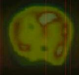
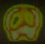
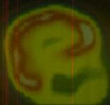
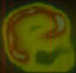
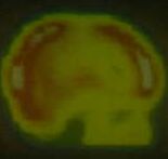
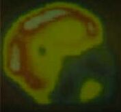
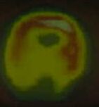
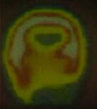
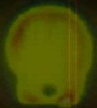
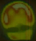
| SPECT or Single Photon Emission Computerized Tomography is technically known as HMPAO brain SPECT study performed after intravenous administration of 20 mCi of Tc-99m HMPAO. In this study, Tc-99, a very short acting approved medicinal quality radioactive chemical is injected into the patient. The dye concentrates in areas with high perfusion. A Gamma camera is used to photograph the part of the body being studied, using the radioactivity of the dye itself. Highly perfused areas show up as whitish to red areas (top part of image shown on the left) while medium perfused areas show a yellow color. Low perfusion areas show a green area while perfusion-less areas show as bluish areas (bottom part of image on left) |
| Name of child and results |
SPECT before HBOT |
SPECT after HBOT |
| Ambar Good perfusion seen to the cerebral parenchyma. Moderate improvement in cerebral perfusion is noted on comparing with pre treatment scan. (Note the small white patches with broken red band forming the outer circular zone in the midst of the yellow background on left (before) and how much of it is changed to white and red and a more complete circle on the right (after), signifying increased perfusion. |
 |
 |
| Arush Good perfusion seen to the cerebral parenchyma. Cerebral perfusion is maintained on comparing with the pretreatment scan. Note the more complete ring of white to red zone on the right (after). |
 |
 |
| Asavari Good perfusion seen to the cerebral parenchyma. Cerebral perfusion is maintained on comparing with the pretreatment scan. Some increase in size of white zone on top part of the brain on right side (after). |
 |
 |
| Ashutosh
Good perfusion seen to the cerebral parenchyma. Moderate improvement in cerebral perfusion is noted on comparing with the pre treatment scan. Note the marked increase in size of white patches of increased perfusion on right (after) |
 |
 |
| Salil Mild hyperperfusion is seen in bilateral occipetal lobe. Tracer distribution in frontal, temporal and parietal lobe is good. Mild improvement in overall perfusion to the cerebral cortex on comparing with the previous study. |
 |
 |
| Tushar Good perfusion seen to the cerebral parenchyma. Moderate improvement in cerebral perfusion is noted on comparing with the pretreatment scan. Note the transition of the broken faint yellowish-red ring to large white to red zones of high perfusion on the right (after) |
 |
 |
| Click here to go to top |